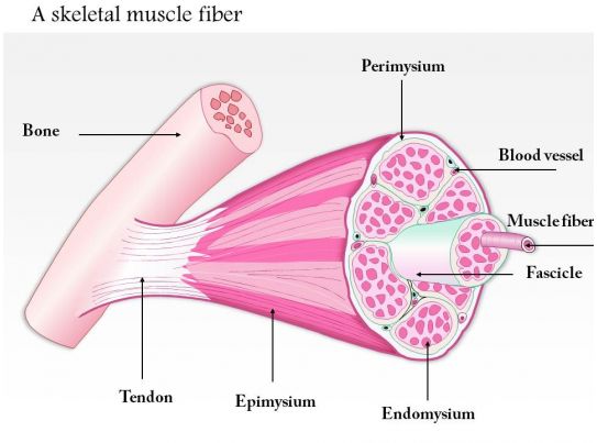
38 muscle fiber model with labels
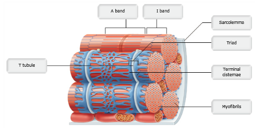
Cardiac Muscle Fiber Model - Spectrum Scientifics This block model depicts a cardiac muscle fiber that has been greatly enlarged to represent significant anatomical features such as the intercalated discs. Also includes detailed representations of sarcolemma, T-tubules, myofilaments, nucleus, and mitochondria. Mounted on a base. Dimensions: 15.75" x 10.25" x 7.5". Muscle Fiber Model Labeled Anatomy Muscle Fiber Model Labeled Anatomy Cardiac muscle fiber model. Sarcomere muscle skeletal bands band anatomy biology structure filament definition structural vs microscope associated muscular physiology isotropy cells below proteins.
Muscle Tissue Diagram Labeled Muscle Tissue Diagram Labeled. The Digestive System | Digestive system, Lpn to rn, Stratified squamous we have 7 Images about The Digestive System | Digestive system, Lpn to rn, Stratified squamous like Print Exercise 14: Microscopic Anatomy and Organization of Skeletal, Skeletal muscle fiber model and also The Digestive System | Digestive ...

Muscle fiber model with labels
Skeletal Muscle Fiber Labeling - Printable This is a free printable worksheet in PDF format and holds a printable version of the quiz Skeletal Muscle Fiber Labeling. By printing out this quiz and taking it with pen and paper creates for a good variation to only playing it online. muscle anatomy not labeled Histology respiratory trachea cartilage labeled perichondrium epithelium hyaline system tissue slides anatomy lamina propria embryology edu label tract labels human. Anatomy 99worksheets. Muscle junction neuromuscular fiber anatomy muscular system muscle fiber model labeling Diagram | Quizlet muscle fiber model labeling STUDY Learn Flashcards Write Spell Test PLAY Match Gravity Created by crlavenuePLUS Terms in this set (7) transverse tubule sarcoplasmic reticulum triad sarcolemma myofibril consists of actin (thin) & myosin (thick) fibrils sarcomere nucleus Subjects Arts and Humanities Languages Math Science Social Science Other
Muscle fiber model with labels. Skeletal Muscle Fiber Model - Myofibrils - YouTube This video was produced to help students of human anatomy at Modesto Junior College study our anatomical models. PDF MUSCLE MODEL ACTIVITY GUIDE - Field Museum muscle model / The Machine inside: BioMechanics activity guide For stud E nt ACTIVITY - Exploring Muscle Size Most animals, including humans, are born with the exact number of muscle fibers they will have their entire lives. This means they cannot increase their density, but they can change the size of muscle fibers. Explore how fiber size ... Muscles Labeling - The Biology Corner Nov 11, 2020 · Muscles Labeling. Shannan Muskopf November 11, 2020. This activity is aligned to my anatomy and physiology curriculum where students study the structure and function of muscle tissues. This has been a challenging topic to cover remotely because I can’t use traditional models. Typically, I would use straws and rubber bands to model fascicles ... General Anatomy of Skeletal Muscle Fibers - GetBodySmart Skeletal Muscle Fiber Location and Arrangement. are located inside muscles, where they are organized into bundles called […] Internal Anatomy of Skeletal Muscle Fibers. An interactive quiz about the internal anatomy of skeletal muscle fibers, featuring illustrations-based multiple choice questions.
Sarcomere Model | Muscle Fiber Model | Skeletal Muscle - 3B Scientific This micro-anatomy model magnifies the anatomy of the human muscle fiber approximately 10,000 times. This muscle model illustrates a section of a skeletal muscle fiber and its neuromuscular end plate. The muscle fiber is the basic element of the diagonally striped skeletal muscle. You've never seen a muscle fiber in this way! This high quality muscle fiber replica brings a hands-on understanding of the human muscle to any classroom. Muscle Fiber Model (Altay) Flashcards | Quizlet Muscle fiber model identifications Terms in this set (21) sarcolemma Identify the membrane. endomysium Identify the tissue layer. myofibril Identify the structure. thick myofilament Identify the structure. thin myofilament Identify the structure. neuromuscular junction Identify the connection. axon Identify the structure. axon terminals Muscle Fiber Micro Anatomy Models - GTSimulators.com We are an authorized dealer of Muscle Fiber Micro-Anatomy Models. We carry also myofibril of striated muscle Models, Skeletal Muscle Fiber Model and Complete Sarcomere Models. Visit us online to buy Muscle Fiber Micro-Anatomy Models on sale. all department; Anatomy Models . Skeleton Models; 3D Printed Anatomy Models; Muscle Fiber Model #1 - Ohio University - YouTube Muscle Fiber Model #1 - Ohio University - Anatomy & Physiology
Muscle Models | Muscle Figures | Musculature Models 3B MICROanatomy™ Human Muscle Fiber Model, 10,000 times magnified - 3B Smart Anatomy. $ 339.00. Item: 1000213 [B60] This micro-anatomy model magnifies the anatomy of the human muscle fiber approximately 10,000 times. This muscle model illustrates a section of a skeletal muscle fiber and its neuromuscular end plate. Skeletal Muscle Slide Labeled - can you name the structures of a ... Skeletal Muscle Slide Labeled - 16 images - how the skeletal system works with the muscular system human body systems, skeletal muscle anatomical features, chapter 7 page 8 histologyolm 4 0, skeletal muscle structure proprofs quiz, anatomy labeled muscle fiber Artery vein histology muscle anatomy muscular smooth arteries microanatomy layers tissue vessel slide blood elastic exam type identify vs vessels. The skeletal muscle fiber stock vector. Microscopic structure of skeletal muscle by dr. s. n. singh anatomy labeled muscle fiber Muscle Fiber Anatomy Quiz - PurposeGames.com This is an online quiz called Muscle Fiber Anatomy. There is a printable worksheet available for download here so you can take the quiz with pen and paper. Your Skills & Rank. Total Points. 0. Get started! Today's Rank--0. Today 's Points. One of us! Game Points. 15. You need to get 100% to score the 15 points available.
Types of Muscle Fibers - YouTube
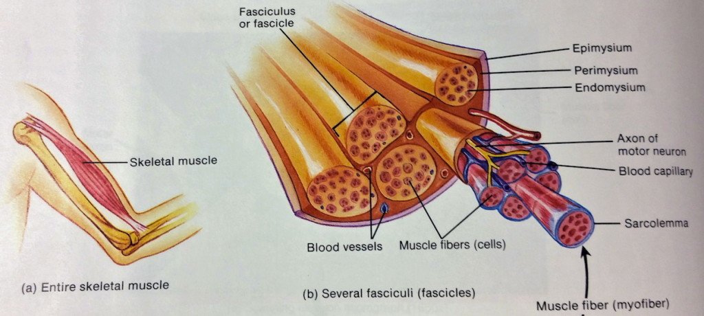
Solved c frame Figure II. Organization of skeletal muscles - Chegg The labels are as follows- 1- Epimysium 2- Muscle fasciculus 3- Nerve fiber 4- E …. View the full answer. Transcribed image text: c frame Figure II. Organization of skeletal muscles Exercise 1: Muscle Organisation Label the connective tissuc layers associated with Figure 11.2. 0 Figure 11.2. Muscle fiber model illustrating connective tissue ...

What Did You Do Today at School?: Muscle Contraction Modeling
muscle fiber diagram labeled Show The Different Between The Three Types Of Muscle Fibers With brainly.in muscles types muscle skeletal fibers three different diagram smooth striated cell body system muscular type involuntary human cells brainly heart Protein actin diagram structure muscle contraction labeled illustration vector illustrations skeletal istockphoto clip.

Skeletal Muscle Histology - Embryology
anatomy labeling muscle anatomy kenhub constrictor muscles ridge medius muscle pharynx pharyngeal musculus middle passavant aspects clinical. Gross Anatomy Of The Muscular System Review Sheet boundbobskryptis.blogspot.com. anatomy. Abdominal Muscle Labeling Quiz . abdominal muscle quiz. Skeletal Muscle Fiber Model . muscle ...

Print Ch. 9 Muscles and Muscle Tissue flashcards | Easy Notecards
Muscle Fibers: Anatomy, Function, and More - Healthline May 12, 2020 · Takeaway. The muscular system works to control the movement of our body and internal organs. Muscle tissue contains something called muscle fibers. Muscle fibers consist of a single muscle cell ...

Muscle Fiber | SpringerLink
Muscle Fiber Model: Motor Neuron, Myeline Sheath, Node of Ranvier ... Dec 20, 2013 - Muscle Fiber Model: Motor Neuron, Myeline Sheath, Node of Ranvier, Synaptic Terminal, Synaptic Cleft, Endomysium, Sarcolemma, Nuclei, Mitochondria, T ...
![Muscle fiber, Skeletal muscle, and Contraction [USMLE] - YouTube](https://i.ytimg.com/vi/35f8Vzd843U/hqdefault.jpg)
Muscle fiber, Skeletal muscle, and Contraction [USMLE] - YouTube
Muscle Model - an overview | ScienceDirect Topics The muscle models have been used in the exoskeleton control schemes. Unlike the dynamic model, the muscle model predicts the muscle forces deployed by the muscles of the human limb joint as a function of muscle neural activities and the joint kinematics (Anam and Al-Jumaily, 2012). The input is the EMG signals and the output is force estimation.
94086359 Style Medical 3 Molecular Cell 1 Piece Powerpoint Presentation Diagram Template Slide ...
skeletal muscle anatomy labeled : Skeletal muscle fiber model, Skeletal muscle anatomy labeling part 1 - YouTube and also Neurolemmocyte On Skeletal Muscle Model - Human Anatomy - GUWS Medical. MRI Musculo-Skeletal Section: MRI Anatomy Of The Shoulder (sagittal View).
The Physiology Of Weight Training - Therapeutic Personal Trainers North Vancouver
Skeletal Muscle Fiber Location and Composition - GetBodySmart A review of skeletal muscle fiber (cell) location, structure, anatomy, and function using interactive animations, models, and labeled diagrams. Start learning now!

http://www.fotoglasswood.blogspot.com / Atau Klick : http://www.pendaftarankursus.wordpress.com ...
SAC A&P Model Key - Muscular System - austincc.edu Muscular System. M1 - Muscled Arm. M2 - Muscle Leg. M3 - Female Muscle Figure. M4 - Microanatomy Muscle Fiber. M5 - Muscle Figure.

32 Label The Structures Of A Skeletal Muscle Fiber - Label Design Ideas 2020
Muscle Fiber. 1. Myofibrils 2. Mitochondrium 3. Postsynaptic membrane 4 ... Muscle Fiber. 1. Myofibrils 2. Mitochondrium 3. Postsynaptic membrane 4. Synaptic gap with basal lamina 5. Presynaptic membrane 6. Presynaptic vesicle 7. Schwann cell 8. Nucleus 9. Actin filament 10. Sarcomere 11. Myosin filament 12. Myelin sheath 13. Neurofibers 14. Cell membrane (sarcolemma) 15. Transverse membrane tube 16. Triad 17.

Altay Anatomical Model Skeletal Muscle Fibre
muscle fiber model labeling Diagram | Quizlet muscle fiber model labeling STUDY Learn Flashcards Write Spell Test PLAY Match Gravity Created by crlavenuePLUS Terms in this set (7) transverse tubule sarcoplasmic reticulum triad sarcolemma myofibril consists of actin (thin) & myosin (thick) fibrils sarcomere nucleus Subjects Arts and Humanities Languages Math Science Social Science Other
32 Label The Structures Of A Skeletal Muscle Fiber - Labels Information List
muscle anatomy not labeled Histology respiratory trachea cartilage labeled perichondrium epithelium hyaline system tissue slides anatomy lamina propria embryology edu label tract labels human. Anatomy 99worksheets. Muscle junction neuromuscular fiber anatomy muscular system

eGFP Expression under UCHL1 Promoter Genetically Labels Corticospinal Motor Neurons and a ...
Skeletal Muscle Fiber Labeling - Printable This is a free printable worksheet in PDF format and holds a printable version of the quiz Skeletal Muscle Fiber Labeling. By printing out this quiz and taking it with pen and paper creates for a good variation to only playing it online.

Muscular System The Neuromuscular Junction - YouTube

V Ling: Art Center Summer Show UPDATE
Post a Comment for "38 muscle fiber model with labels"